X-ray Dose DataSheet 2017.pdf
T h i s A p p l i c a t i o n N o t e s u m m a r i z e s X - r a y r a d i a t i o n d o s e , a n d t h e t e c h n i q u e s t h a t c a n b e e m p l o y e d t o re d u c e r a d i a t i o n e x p o s u re t o s a m …
This Application Note summarizes
X-ray radiation dose, and the
techniques that can be employed to
reduce radiation exposure to samples
in your Quadra
TM
X-ray Inspection
System.
Quadra X-ray geometry
First we need to consider how the Quadra X-ray
Inspection system works.
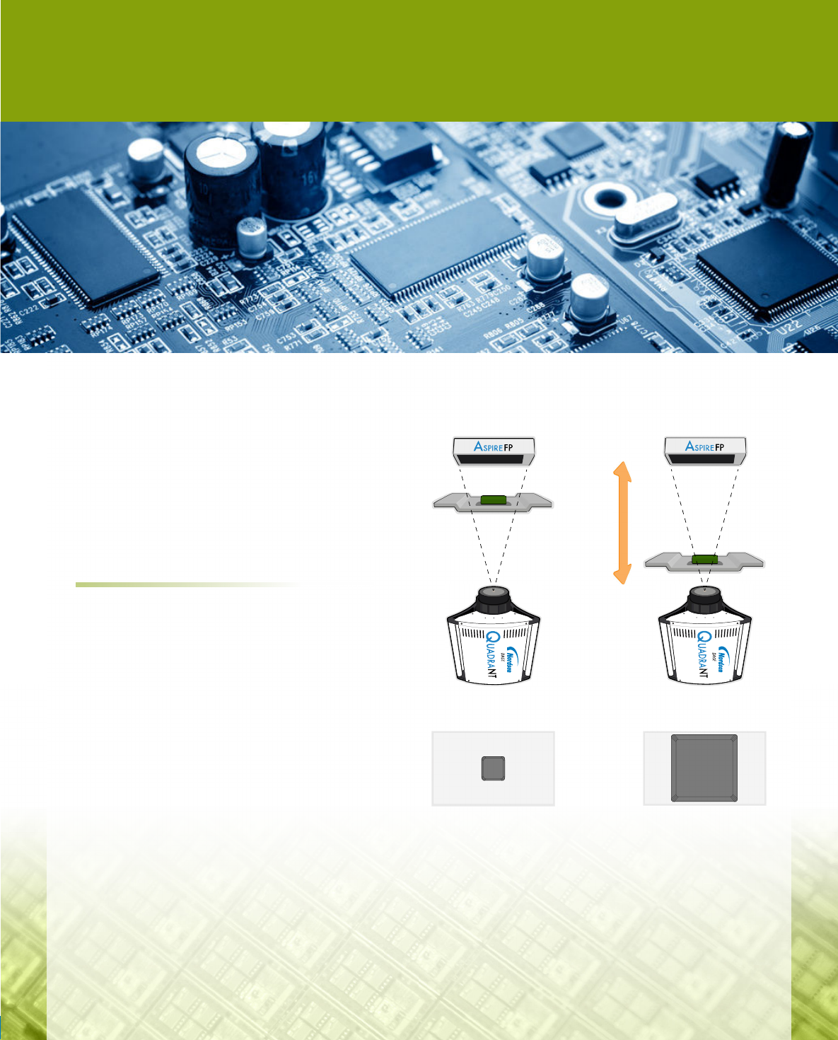
Quadra allows the internal structure of samples to be
seen by shining X-ray light through a sample (figure 1).
This creates a shadow image that is detected in real-
time using a high resolution Aspire
TM
Flat Panel X-ray
detector. The darkness of the shadow cast by any point
in the sample depends on how much X-ray light has been
absorbed by that part of the sample.
High image magnification is used to see smaller
features clearly. This is achieved by moving the sample
closer to the X-ray tube. For the highest magnification,
the sample tray is lowered to the top of the X-ray tube.
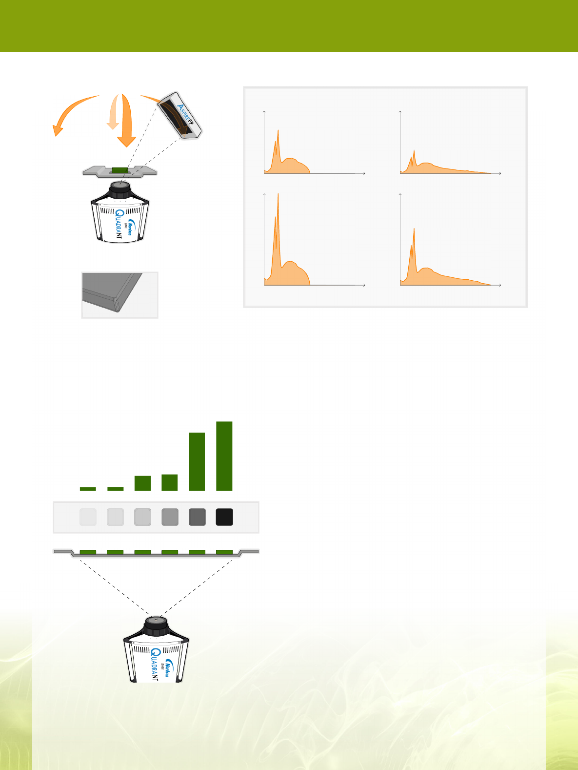
Viewing the sample from different angles allows more
defects to be observed. The Aspire Flat Panel detector
moves around two oblique axes to allow this. X-rays
from the QuadraNT
TM
X-ray tube are emitted over a
wide cone angle so there are plenty of X-rays available
off axis.
Application Note
X
-RAY DOSE CONSIDERATIONS
Low magnification
High magnification
Figure 1: Quadra X-ray tube, sample and detector
geometryshownatlowmagnication(left)andhighimage
magnication(right).
X
-RAY DOSE CONSIDERATIONS
Application Note
Oblique view
SnZnCuSiAl Pb
Absorption
Differentmaterialsabsorbradiationtodifferingdegrees(gure
3). Lead is very good at absorbing X-ray radiation which is
why it’s used to shield the X-ray cabinet. Silicon and aluminium
are relatively poor at absorbing X-rays so images tend to be
very bright. Tin and copper, commonly used in solder, are
somewhere in between.
The shadow created at any point also depends on the voltage
andpowerusedtodrivetheX-raytube(gure4).
An X-ray tube operating at a voltage of 80 kV creates X-rays
over the entire energy range upto 80 keV.
Increasing the voltage to 160 kV doubles the X-ray output
energy range from 80 keV to 160 keV.
Increasing the tube power increases the number of X-rays
emitted at every point over the range.
In general, higher energy X-rays are more likely to pass through
a sample than lower energies. High energy beams are useful
for looking at thick samples, or materials that strongly absorb
X-rays, for example lead.
Lower energy X-rays are better for thinner samples, or
materials that weakly absorb X-rays, for example copper or
silicon.
10 W
0 80 160
Output
keV
0 80 160
Output
keV
20 W
0 80 160
keV
0 80 160
keV
80 Kv 160 Kv
Figure 3: X-ray absorption by different materials of the
same thickness.
Figure 4: X-ray photon energy output for different tube voltage and power settings.Figure 2: Oblique viewing angles
are achieved by moving the
detector in a hemisphere around
the sample.
X
-RAY DOSE CONSIDERATIONS
Application Note
0
40
80
I
keV
Contribution to X-ray image
0 40
80% reduction
80
I
keV
X-ray beam after Zn filtering
100%20%
SiSi
Zn filter
Si Dose
Image
What is X-ray dose?
The X-ray dose for any part of the sample is a measure of
the X-ray energy absorbed per unit mass, and is measured
inunitsofgray(Gy).1Gy=1J/kg=100rad.
Since different materials absorb incoming X-rays with
differingefciency,theirabsorbeddosewillvarywhen
placed in the same X-ray beam. A material like lead which
absorbs X-rays well and casts a dark shadow in an image
will absorb a higher dose than a material like silicon which
absorbs X-rays weakly.
The absorbed dose also varies with:
• Distance to the X-ray source: The amount of X-ray
radiationreducesas1/r
2
in the same way a light appears
brighter the closer you are to it.
• Time in X-ray source: X-ray dose is cumulative so the
longer spent in the X-ray beam, the higher the absorbed
dose.
• X-ray tube output: The higher the tube power or
voltage, the larger the absorbed dose.
How can I reduce sample dose?
Reducingthemagnicationusedandminimizingthetime
the X-ray tube is switched on are effective ways to reduce
X-ray dose. Quadra’s Low Dose mode can be used to
switch X-rays off when it detects the sample is no longer
being manipulated.
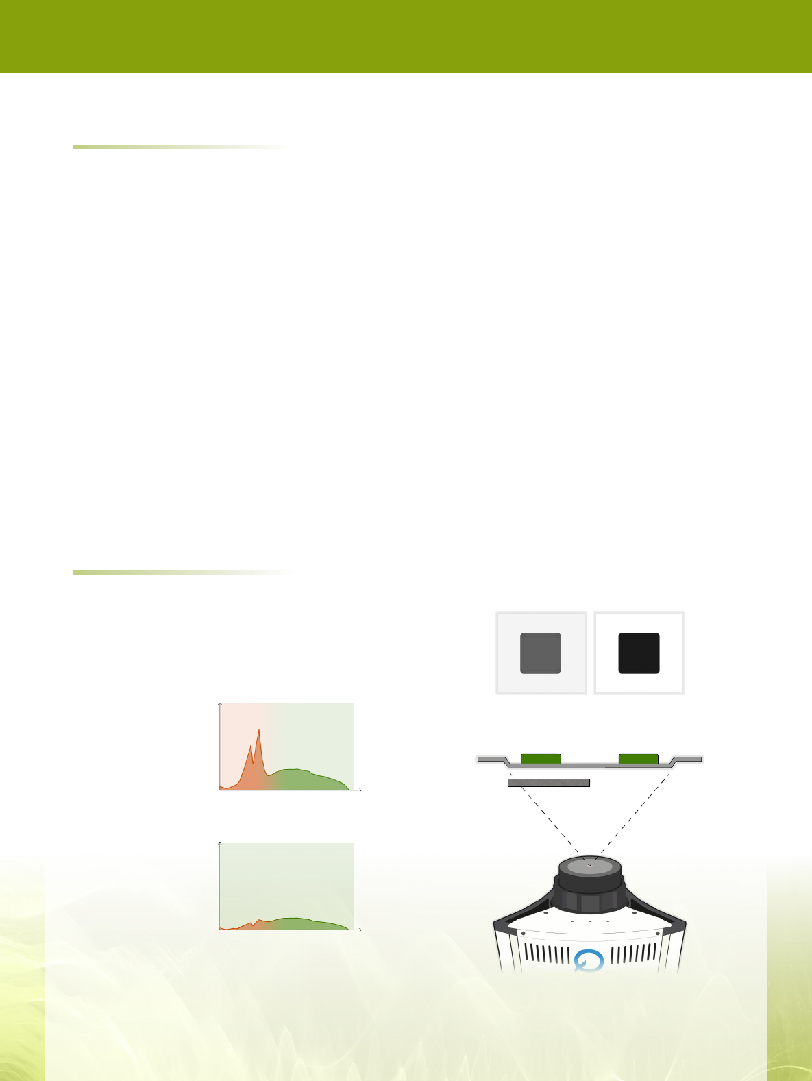
Figure 5: X-ray photon energy output without (top) and with
(bottom)additionalltering.Theenergiesbelow30keV
donotcontributetothenalimagebutstillcontributeto
absorbed dose.
Figure 6: Absorbed dose and image quality for a typical
siliconsamplewithlteringtray(left)andwithout(right).
AnothermethodistolteroutlowerenergyX-rays(gure
5).TheseX-raysdonotcontributetothenalimagefortwo
reasons:
• Absorption by sample: At low energies, particularly
below 20keV, X-rays are strongly absorbed by most
samples. They contribute to sample dose, but are less
likely to make it through the sample to the X-ray detector.
• Detector sensitivity: Flat panel X-ray detectors, like
AspireFP
TM
, are insensitive to X-rays below ~20 keV. Any
X-rays that manage to pass through the sample have a
lower likelihood of being detected.
Filtering out low energy X-rays is an effective way to reduce
sample dose without compromising image quality.
• Alteringsampletray is available for Quadra
TM
which
incorporates zinc strips to absorb low energy X-rays while
letting higher powers pass through to the sample. This
canreduceoverallX-raydosebyupto80%(gure6).
• Masking is a simple way of reducing X-ray dose for any
components on a board that do not need to be inspected
at all by X-ray. Attach a portion of dense material, for
example lead, to the sample tray immediately below the
sample to mask the sensitive component from X-rays.