Cougar_EVO_Brochure_en-LR.pdf - 第6页
ONE CLICK PHILOSOPHY One-click solutions make it easy to perform the advanced manipulations required for fast and reliable X-ray inspec- tion. Such as: - Click & Center - Frame & Zoom - PowerDrive - Zoom+ These f…
The inspection of electronic components during research
and development are complex and need the broadest
range of features and state-of-the-art technology.
Computed Tomography is a must for detailed analyses
of micro components such as those which are used in
batteries, connectors and medical devices.
The new Cougar EVO Plus offers ultimate image resolu-
tion and highest industry CT reconstructions in addition to
other empowering pros:
EXCEPTIONAL CT QUALITY
- The new Panel 1616 detector enables CT volumes
of the highest quality
- Excellent contrast-to-noise ratio
- Highly sensitive detector
VISUALIZATION BY YXLON FF CT SOFTWARE
- Integrated workflow in the FGUI user interface
- Realistic, vivid visualization due to individual 3D
cinematic renderers and a preset selection of transfer
functions (TF)
- The visualization of laminographic volumes have the
same high quality as CT volumes, which are much more
complex
- Artifact reduction such as BHR Beam Hardening
Reduction, BHC Beam Hardening Correction, Ring
Artifact Reduction, Noise Reduction Volume, etc.
- Clear visual fault detection
ADDITIONAL PROS
Available as an option
- New detector Panel 1616:
· High speed
· Consistent image quality thanks to the stable
detector temperature
· No impact of radiation on lifetime (radiation resistant)
Laboratory inspections:
leading technology for precise analysis
APPLICATIONS
- Batteries
- Connectors
- Various hard-to-see electronics components
- Medical material
- Military and space electronics
5
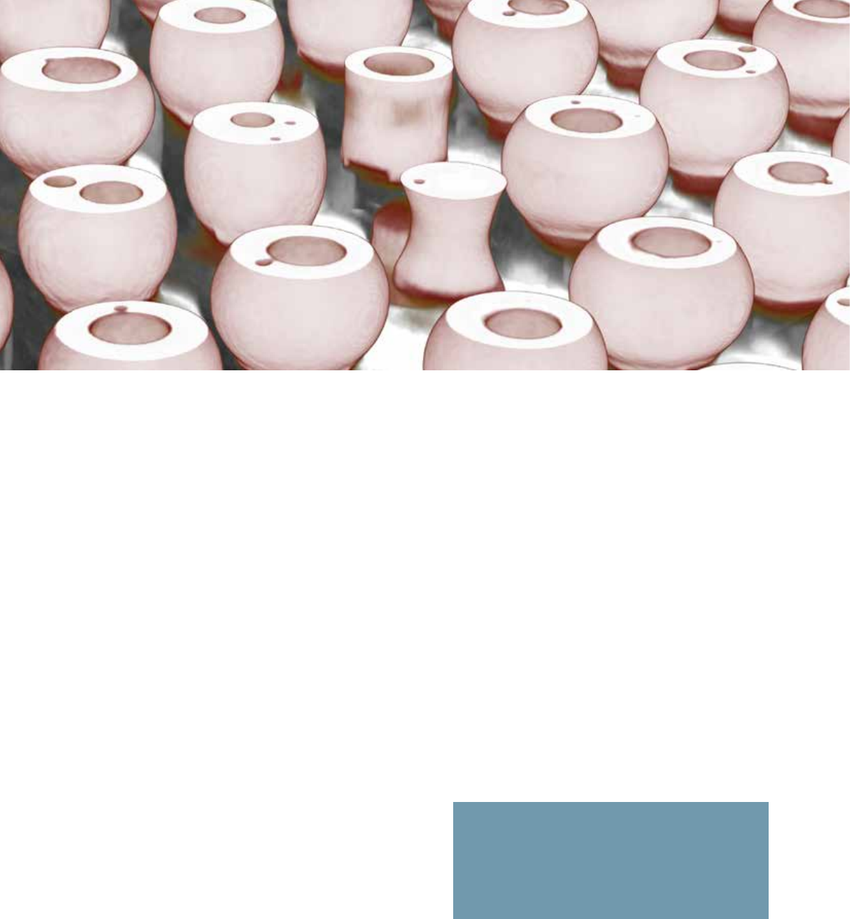
3D Laminography image Ball escaped in Via
ONE CLICK PHILOSOPHY
One-click solutions make it easy to perform the advanced
manipulations required for fast and reliable X-ray inspec-
tion. Such as:
- Click & Center
- Frame & Zoom
- PowerDrive
- Zoom+
These functions guarantee constant-intensity magnifica-
tion without tube adjustments or software interpolation,
and can be carried out with one simple click.
EXTENDED BGA INSPECTION
With Cougar EVO, you can quickly select and index
individual balls, either manually or using automatic grid
detection. A wizard guides you step-by-step through the
workflow and ensures perfect accurate and repeatable
results. Plus, the feature allows multiple operators to run
the same inspection routines.
EXTENDED ADR INTERFACE
Cougar EVO software can be tailored to individual
requirements, with operators free to define their own
specific analysis. This also includes customized algo-
rithms.
eHDR-INSPECT
To ensure highest product quality, the eHDR filter
highlights complex structures with just one click. Thanks
to our advanced software and enhanced 16-bit gray scale
values, it detects even the slightest variances in gray
scale, so that no defect will be missed. This allows you
to easily see faults that were invisible before.
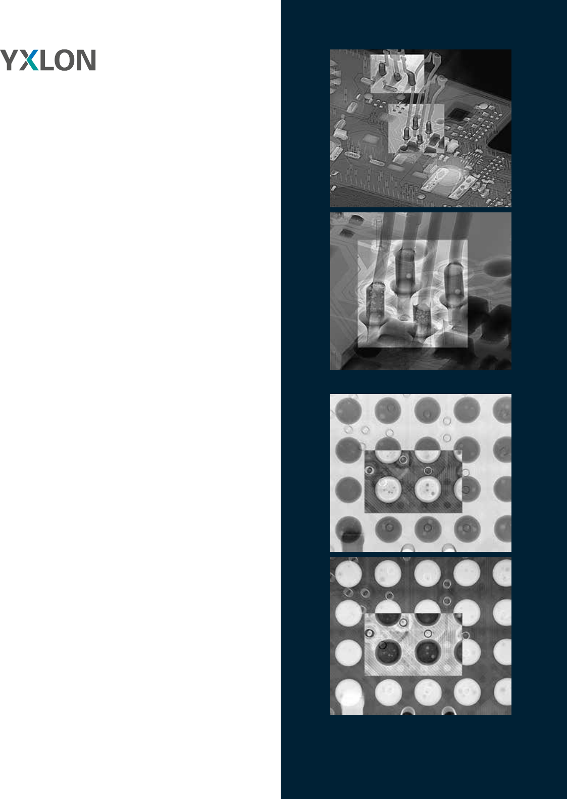
2D MULTI AREA VOID CALCULATION (MAVC)
QFNs and other bottom-terminated devices without
overlapping can also be inspected with 2D digital
radioscopy. Faulty or missing solder joints and large areas
of voiding are reliably detected, and MAVC helps analyze
voids in complex soldering designs. With just four
parameters to adjust, setup is quick, simple, and cost-
efficient. Precise void analysis of multi-layer components
needs computed laminography and VoidInspect.
Functions that
bring you forward
6
Using eHDR for Regions of Interest ROI with details (voiding in a BGA ball)
THT with voiding
77
YXLON COUGAR EVO
Check out these facts
X-ray inspection system
Dimensions (w x d x h) 1,000 x 1,050 x 2,200 mm
Weight 1,450 kg
Mains connections
230 V ± 10% AC, 50/60 Hz, 1 Phase,
neutral and ground conductor
Fuse protection 16 A
Max. power consumption 2.5 kVA
Max. dose rate* < 1µSv/h
* at 100 mm distance to the cabinet surface
Inspection parts
Max. part size 440 mm x 550 mm (17” x 21”)
Max. radiographic area 310 mm x 310 mm (12” x 12”)
Max. part weight (standard) 5 kg
Max. part weight rotation and tilt 2 kg
X-ray source FXT-160.50 Microfocus FXT-160.51 Multifocus
Target transmission
Voltage range 20 – 160 kV
Current range 0.001 – 1.0 mA
Tube power max. 64 W
Target power max. 15 W
Target material Tungsten
Detail detectability 0.75 µm < 0.3 µm
X-ray intensity control TXI
Image Chain
Geometric magnification ~ 2,000 x
Total magnification ~ 256,000 x
Spatial Resolution 1.5 µm 0.6 µm
General Product Features
Time to first image (typ.) ~ 10 s
Reconfiguration time (typ.) < 60 s
Acquisition time (Quick Scan)
for 2000 projections
~ 3.15 min
Reconstruction time (Quick Scan)
for 2000 projections
~ 1.55 min
Acquisition time (micro3Dslice
Semicon) for 120 projections
~ 1.45 min
Reconstruction time (micro3Dslice
Semicon) for 120 projections
~ 0.30 min
Access for sample loading manual
Cabinet window 380 mm x 200 mm
Monitor 27” Ultrasharp, wide viewing angles
Zoom+ yes
Manipulation
Manipulation control via mouse or joystick
Manipulation axes X, Y, Z(D)*
Oblique viewing +/-70° (140°)
* Manipulation options for horizontal and vertical rotation and tilting available
Detector Y.Panel 1308 Y.Panel 1313 ORYX 1616
Max. resolution Pixel 1004 x 620 1004 x 1004 1276 x 1276
Pixel size 127 µm²
Pixel area 128 mm x 79 mm 128 mm x 128 mm 162 mm x 162 mm
A/D transformer 16 bit
Please note that not all components and features described in this brochure belong to the standard configurations but are part of an optional selection.