semi合集-English.pdf - 第7285页
SEMI MF847-0705 © SEMI 2003, 2005 5 10 Procedure 10.1 Position the detector so that the ang le between the extensio n of the incident X-ray beam and th e line joining the detector and the axis of rotation of the specim e…
SEMI MF847-0705 © SEMI 2003, 2005 4
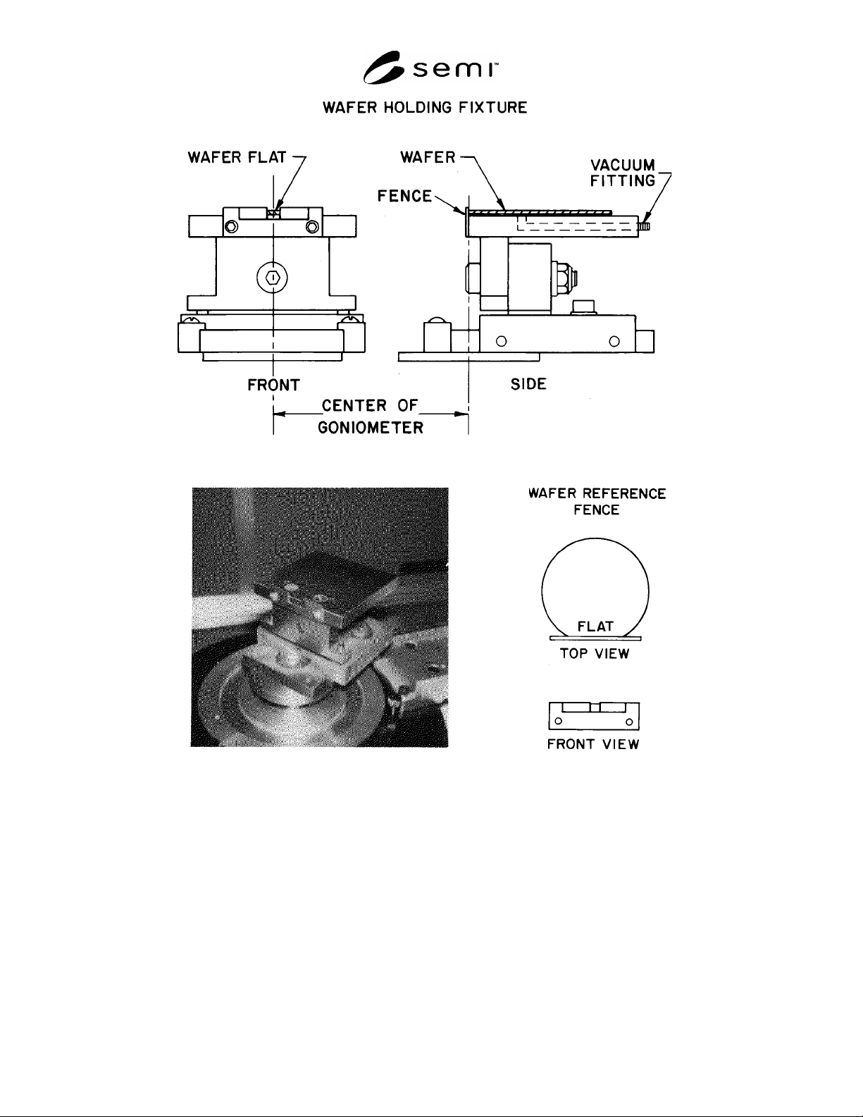
a. Front and Side Views
b. Photograph of Mounted Fixture c. Detail of Wafer and Reference Fence
Figure 1
Wafer Holding Fixture for X-Ray Edge Diffraction Method
9.2.1 The horizontal surface must be parallel to the plane of the X-ray beam so that the diffracted beam impinges on
the detector (see Figure 2).
9.2.2 Both the side of the reference fence against which the wafer flat is located, and the fixture surface to which
the reference fence mates, must be flat to within one part in 10,000.
SEMI MF847-0705 © SEMI 2003, 2005 5
10 Procedure
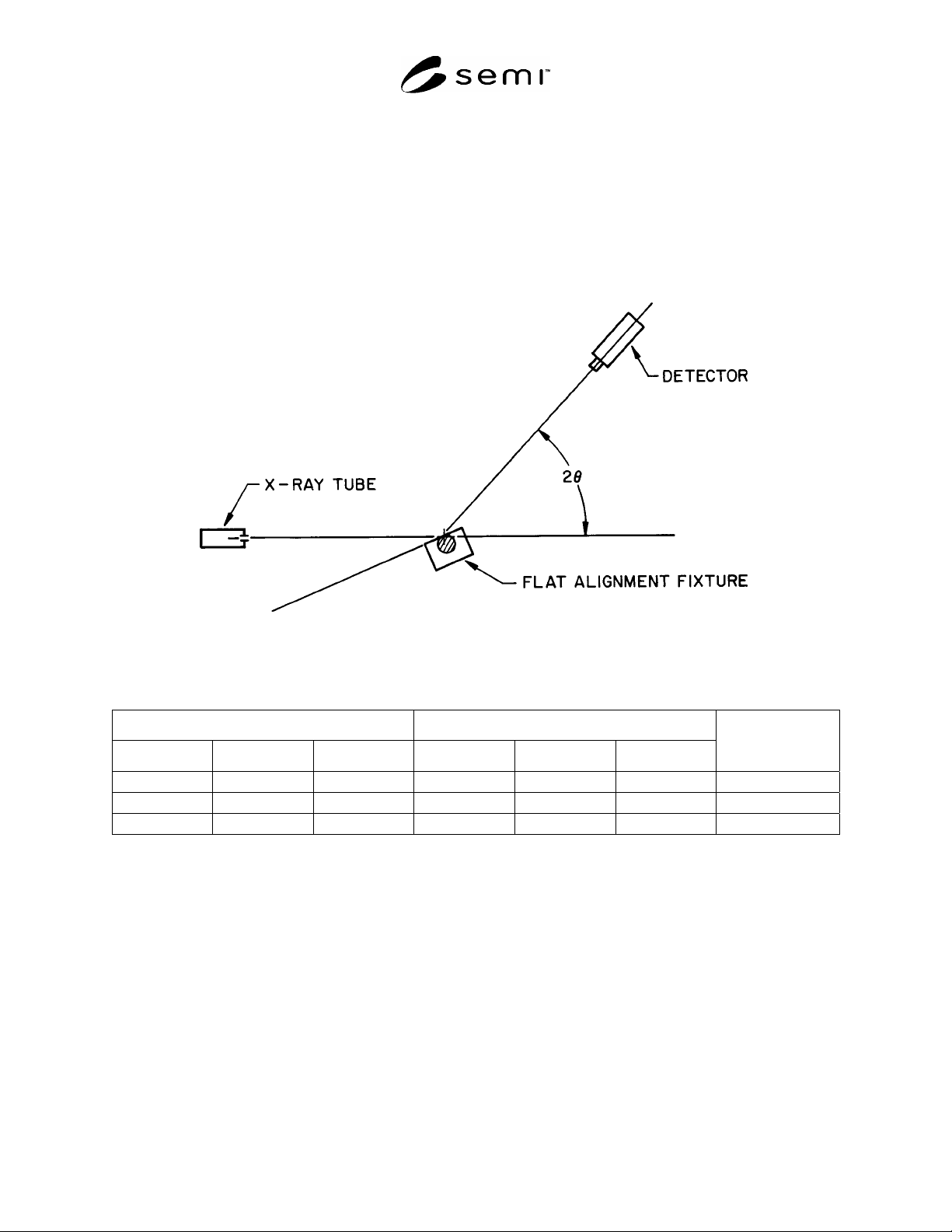
10.1 Position the detector so that the angle between the extension of the incident X-ray beam and the line joining
the detector and the axis of rotation of the specimen is equal (to the nearest minute) to twice the Bragg angle
(see Figure 2).
NOTE 3: This angle (twice the Bragg angle) is listed in Table 1 for CuK radiation for the recommended reflecting planes
corresponding to common silicon wafer flat locations.
10.2 Place the wafer to be tested on the fixture, front surface up. Take care to ensure that the flat is securely located
against the reference fence and activate the vacuum holddown.
Figure 2
Schematic of the Diffraction Geometry for the X-Ray Edge Diffraction Method
Table 1 Bragg Angles,
, for X-Ray Diffraction of Cu-K Radiation in Silicon Crystal
#1
Flat Location Recommended Reflecting Plane
h
k l h k l
Detector Location (2
times Bragg Angle)
1 1 0 2 2 0
4720
2 1 1 4 2 2
8808
1 0 0 4 0 0
6912
#1
Wavelength
= 1.5417 Å.
10.3 With the goniometer movement mechanism, adjust the fixture about the axis of rotation perpendicular to the
incident and reflected beams until the diffracted intensity is at a maximum.
10.4 Record to the nearest 1 min, as
1
, the angle that is indicated on the goniometer.
10.5 Remove the wafer from the fixture, turn it over so that the front surface is now down. Again, take care to
ensure that the flat is securely located against the reference fence and activate the vacuum holddown.
10.6 With the goniometer movement mechanism, adjust the fixture about the axis of rotation perpendicular to the
incident and reflected beams until the diffracted intensity is at a maximum.
10.7 Record to the nearest 1 min, as
3
, the angle that is indicated on the goniometer.

SEMI MF847-0705 © SEMI 2003, 2005 6
11 Calculation
11.1 Calculate and record the average angular deviation as follows:
2
31
(1)
where:
= average angular deviation,
1
= first angle reading taken on goniometer, and
3
= second angle reading taken on goniometer.
12 Report
12.1 Report the following information:
12.1.1 Identity of samples tested including supplier and supplier lot identity,
12.1.2 Date of test and identity of operator making the measurements,
12.1.3 Specified flat and surface orientations, and
12.1.4 Measured values of
1
and
3
and the calculated value of
for each wafer.
13 Precision and Bias
13.1 The single-instrument, single-operator repeatability of this measurement was estimated by measuring one
wafer 50 times (25 times each side). This test yielded a distribution of calculated values of
with a 1-
value of
1.94 min.
TEST METHOD B — LAUE BACK REFLECTION X-RAY METHOD
14 Summary of Test Method
14.1 In this test method the wafer is mounted in a Laue back-reflection X-ray camera and a collimated beam of
“white” (continuous or Bremsstrahlung) radiation is directed at the wafer flat.
14.2 A spot is produced on the film for each set of crystal planes that satisfies the Bragg equation for any
wavelength component of the impinging radiation.
14.3 The pattern on the film is read with an engineering drafting head.
14.4 When the flat surface is within 5° of the specified low-index plane, the angle between the nearest zone of Laue
spots that goes through the center of the pattern and the zero reference line is a direct measure of the angular
deviation.
15 Apparatus
15.1 Commercially Available X-Ray Diffraction Apparatus — Utilizing a silver or tungsten tube as the X-ray source
and including a shutter to control the X-ray exposure.
15.2 Laue Back-Reflection X-ray Camera — With the following features (see Figure 3):
15.2.1 Mounting Track — With the upper surface and one side round precision flat perpendicular to each other,
aligned with the X-ray beam from the source.
15.2.2 Wafer Holding Fixture — On which two plane surfaces are ground so that when it is clamped to the
mounting track one surface is perpendicular and the other is parallel with the horizontal (upper) surface of the
mounting track to 1 min of arc (29 m in 100 mm) (see Figure 4). The vertical surface contains holes connected to a
vacuum line through a fitting on the back of the fixture. In use, the flat is aligned to the horizontal reference surface
and the vacuum holds the wafer against the vertical surface.